Blender for Molecular Biology
Describing functions of biological macromolecules are demonstrated by computer animation using 3D CG software Blender. Here the free software Blender allows smooth exchange of the animation scripts developed among users. There are blender script files and the parts of the molecular objects are demonstrated. Although these models and scene graphics needs to be improved for the production uses in animation studios, this development project is ongoing to provide the parts of molecular animation.
A Model of Working RNA Polymelase
An animation model for RNA polmerase that generate mRNA polymer chains from DNA chain was studied. Based on the modeling results published with atomic model data in PDB motions of nucleic acid chains was constructed. Structural Basis for the Transition from Initiation to Elongation Transcription in T7 RNA Polymerase. Yin, Y.W., Steitz, T.A.(2002) Science 298: 1387-1395
file
- blender script rnapol75.blend
- PDB model 1msw.pdb
- ribbon model in X3D 1msw-p.x3d
- DNA and RNA model in X3D 1msw-na.x3d
- saved media rnapol75.mp4
There are method to move nucleic acid polymer along the helical path with Blender. In this case Lattice Modifier was applied because it is useful to control directions of nucleic acid base which should be paired to the corresponging template. Three Lattice objects for the RNA and DNA double helix was made from the PDB coordidnate by a hand work. Array of nucleic acid residues are placed on an Z axis and motion along Z axis is applied. Then this rows of residues are constrained to move in the Lattice. This method has limitation of Lattice length (maximum 64), and we may probably need to take different method eventually.
Modeling Molecular Interaction of Actin and Myosin
The thick filament and thin filament in the skeletal muscle are placed on the scene to examine interact by the motor protein molecules myosin to the actin on the thin filament. The sliding of two filaments may be caused by the myosin head. The laver-arm hypothesis explains the movements are considered to provide a force to pull a thin filament. The model shows an example of such a motion where the interactions between myosin head on the actin polymers. Using these models, we may also examine the long distance walk of myosin head may also examine on this model.
The myosin heads are placed at 14.3nm interval where molecular interactions of them are not yet known, however a tentative laver-arm motions are applied in the animation script. PDB data 1ATN for actin (rabbit skeletal) polymerized as Holmes model, 2MYS for the myosin head (chicken cardiac) are depicted as surface model using UCSF Chimera, then saved as polygon mesh data in STL file format. The thick filament model was imported by EMDatabase #1950. The embedded myosin heads are tentatively removed from the volume by hand works, using the Volume Erase function in UCSF Chimera. ... Tools -> Volume Data -> Volume Erasor)
Tredmilling of Actin
The actin filament moves in the cell by tredmilling that actin polymerize one end and depolymerize on the other end. Taking advantage of the particle method function in Blender, this primitive model demonstrate the random suspension of actin monomers are driven to the filament. In this animation, all actin monomers approached will align to fit the helical formation, although it would never happen in nature. Thus we may need to introduce some of actins failed to polymerize and dropped out from the filaments.
Blender version 2.5 introduced a major improvement in the particle system which allows object move by physics simulation. The actin filament is made with 78 helical arrays of rectangles, where the "emitter" mode is used to locate actin on the helix, taking "emit form" as "Faces" at the same number of emission 78. The times for the emission start and stop are controlled to give a good elongation cycle. There is "keyed" option in the Physics tab in the object property sheet, the emitted particle target are specified. Two big rectangle planes are used as the targets so that particle traverses to the other end of the filament. This is a chain of the particle emitter in Blender specifying beginning and ending time. In case the motions are not always satisfactory for the use in other animation script, we may convert these motions into the sequence of key frame animation..
In Vitro Motility Assay
The in vitro experimental assay is useful to observe motility of kinesin molecules fixed on a plate that allows microtubule filaments to slide on the plate. The motions of microtubules are tracked by attached fluorescence by the microscope. This animation picture demonstrates how the molecules reside in this plate. The motion of microtubule is in the predefined path because a realistic simulation for the motility is not available.
file
- blender script view-ivma10.blend
- a tublin texture on a cylinder mt13sidetop1.png
- texture mapping work on Blender mt13-3ab2-texture.blend
- saved media ivma10.mpg
This microtubule is a flexible cylinder mesh with texture mapping display of the tubulin monomers, so that a lot of microtubules may be introduced. The texture mapping procedure in Blender was saved in the script file that can also export as a different polygon mesh model format.
A Closer Look at Kinesin and Microtuble Interaction
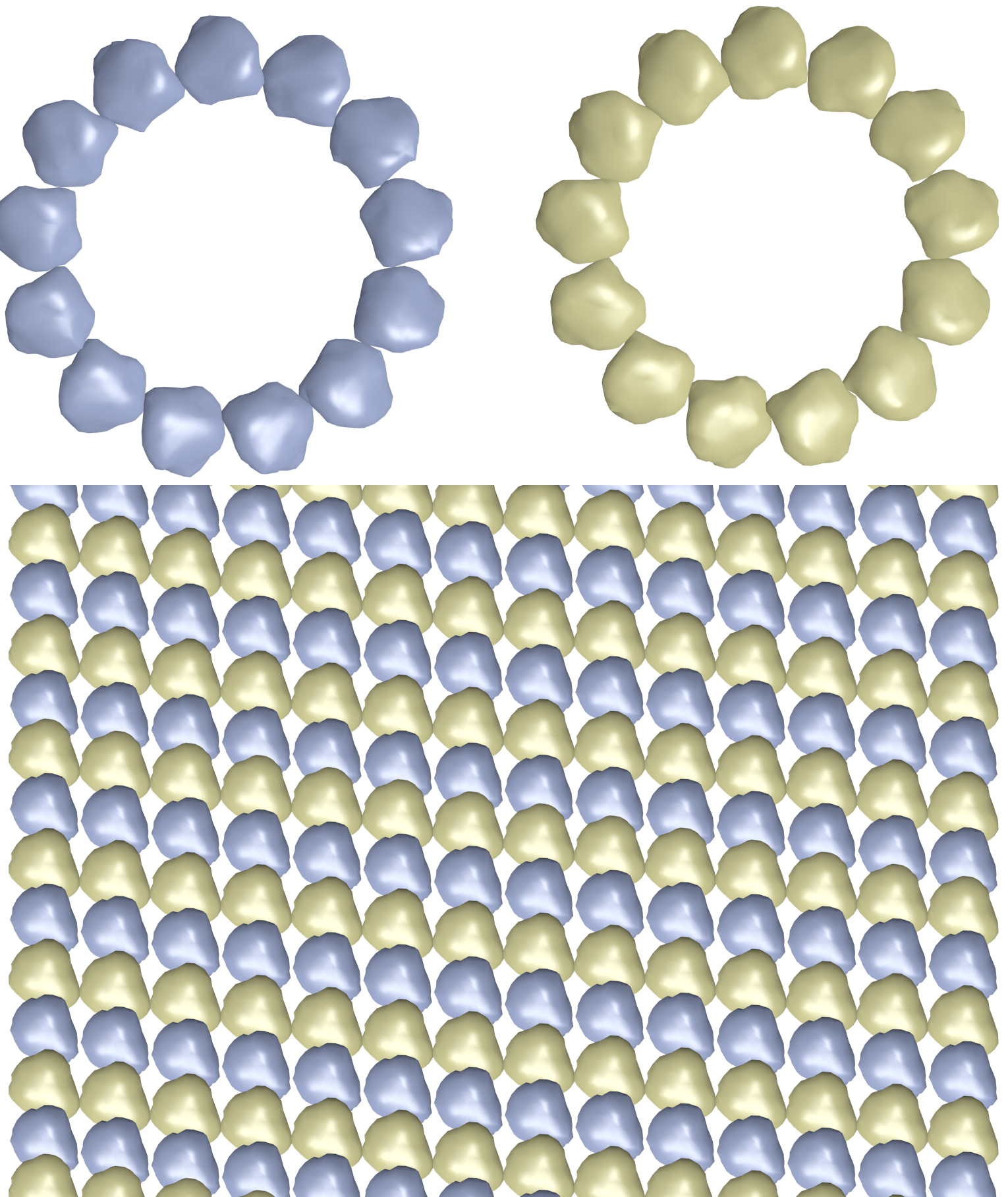
On a hypothetical model of kinesin bound to a microtubule filament this movie demonstrates a camera work to look into the molecular interaction of these proteins. The Microtubule made up with alpha and beta tubulin monomers, colored in yellow and purple, in a helical arrangement.
PDB data 3KIN atomic coordinates only provides 372 amino acid residues, so that tail regions in an alpha helix coiled-coil was inserted from other protein, tropomyosin 1CGA PDB data, additional 89 residues aligned by additional 8 residues overlap, and resulting 17 nm in length. The polygon mesh file in OBJ format was used.
A Theoretical Model of an Artificial Muscle
An application of muscular proteins for the motility system has been studied molecular robot. Dr.Shin-ichro M. Nomura at Tohoku University reported possible construcion of an artificial cell yielding motitliy wby motor proteins. This animation demonstrates how the motor proteins work inside the molecular robot. Here only a few microtuble filaments are depicted that moves by kinesion motor pushing each other filaments toward outer membrane of the cell.
Kinesin and microtubles are the same as previous example.
A working model of Alpha-Amylase
A hypothetical working model of alpha-amylase, the enzyme for the hydration of starch. A hypothetical working model of alpha-amylase, the enzyme for the hydration of starch is demonstrated. An amylose, a chain with 10 glucose monomers, approaches to the active site of the alpha-amylase and divided into some maltose molecules in a raw. The path was predefined from handworks, because it was not yet known how the molecule recruits this substrate into the active site. The thermal vibration of the amylase is introduced by a composition of the vibrational mode from the normal mode analysis. The amplitude was 5 times larger than the room temperature behavior to exaggerate the movements. The active site of the enzyme stays in an original conformation, while the loop regions move a lot suggesting some help for the work of this enzyme.
The ribbon model from the PDB data 1AMY is saved as STL polygon mesh format, and imported by Blender. Ball and stick model of amylose is made by Bioblender, an add-on software for Blender, 3'and 5' oxgen of maltose is colored in pink and red at different material type, they are overlapped in the connected chain. In Blender, "follow path" modifier is used to follow the chain along a freehand path of Bezier curve, at the constraint parameter 0.5. Each maltose was dispatched from the active site using key frame pose with rotation and location on the time line editor.
The catalytic active site of alpha-amylase
The atomic model is animated based on the PDB coordinates data and the in between structures are interpolated. The ribbon model and ball-and-stick model were generated using UCSF chimera and saved as X3D polygon models. Using models of confomational change for the atomic coordinates of the protein were reported at EzCatDB Enzyme database. Blender provides functions for this morphing animation between these different PDB data to describe the chemical reaction of the enzyme. In addition, dotted lines are added to indicate the covalent bond interactions.
file
- blender script
ez1amy-t3e5g.blend
- atomic coodinates data u1amy.pdb.gz
- X3D polygonn model u1amy2.x3d.gz
- Lua program to make skeletal motion data ezc2bvh4-1amy.lua
- skeletal motion data ez1amyb4.bvh
- saved media ez1amy-t3e5g.mp4
- atomic coodinates data u1amy.pdb.gz
THe atomic models for the reaction intermediates are provided from EzCatDB. The motion data of reacting atoms are converted into the BVH motion data. Then the inported polygon model for the protein including reactin side chains and regand molecules are assigned motions. Each primitive for ball and stick models are asigned its singe parent bones. In many case, X3D polygon model file does not come with the chemical information of the protein, so that this script also read the reference PDB file data and set the residue and atom information to the graphics primitive by checking atomic coorinates.
Tips for Blender
Tiling Windows
When working on the multiple windows or panels tiled inside Blender, there are nice ways to handle the triangle icon. First, if you drag the digonal corner of this triangle outside the tile, the tiled window is marged to the adjacent tile and disappears. The adjacent tile have to be single tiled. These are directions out of the two base of isoscales right-triangle. Second, if drag inside to the tile, new tile is created. It is a in-bound direction of the hypotenuse. Right clock menu may not be useful for tiling although the official wiki of Blender advices.
Duplicating Objects without Data
Sometimes duplicated objects without data does not act correctly with bone animation. In that case making the objects real works well.
Working on Big Model
In general, when you place more than 1000 objects, Blender becomes difficult to operate. Even if I add more memory or change the graphics card, it doesn't help much. In particular, ball-stick models and space-filling models tend to be heavy, as atomic models of proteins contain several thousand atoms. Additionally, extensions such as Bioblender often take a very long time to load. It seems to be unavoidable when we create objects using Blender's API. However, once you save it to a Blender file, it will load fast, but the overall operation will remain slow. Since a sphere is displayed as a polyhedron, it will be somewhat lighter if it is made into an octahedron. Many complex polygonal models can be loaded and manipulated relatively quickly, so ribbon models are no problem. For example, when importing the hemoglobin tetramer of PDB model 1A00, there are approximately 4000 atoms, and it is difficult to handle a model in which all atoms are represented as spheres. A model with only the α carbon has 794 residues, which is the usable range. On the other hand, the ribbon model had 280,000 polygons and consumed about 56MB of memory, but it did not become heavy.
Mesh deformation along a Curve
Blender has several different methods, but two methods are useful. If there is no deformation along the curve, either FollowCurve of the constraint, or Deform-Curve of the modifier if there is deformation. In both cases, the parallel movement motion is placed on a curve, so when inserting motion with a keyframe, it is not possible to specify the desired position while looking at the curve. Specify with a variable. For example, if you assign the x-axis to a curve, change the object's transform value and move it to the desired position. At this time, the y and z components may deviate from the curve, causing the mesh to become distorted. Also, it seemed easy to change the orientation of the object by changing the normal of the curve. If you try to directly manipulate an object, the orientation is different from that on the screen, so it is difficult to manipulate the object in the desired orientation.
Camera works
Blender documents explain a method to place the camera on a specified curve and take a picture of the point of interest, but adding constraints to the path will make it troublesome, so just specify a few points as key frames. That was enough. In this case, you can change the point of interest or angle with each keyframe. You can change it naturally by selecting the camera as an object, displaying the camera view, and moving with the G key or rotating with the R key.
Movie File Conversion
Among variations of video format conversion tools available, ffmpeg was mainly used because of rich functions and low problem rate. In our development days, mpeg2 format was useful, however, currently mpeg4 format at specific codec is nice to take.
Others
Working Environment
- blender
- ver 3.0 and 2.70a, windows7 64bit
- UCSF chimera
- ver 1.8 (build 38824), 2013, windows7 64bit
- PC
- windows7 64bit, core-i7-3770S (3.10GHz) 8GB
Acknowledgments
This work was supported by JSPS KAKENHI Grant Number 20K12072 and 24104004. The software was developed at a research institute, National Institute of Advanced Industrial Science and Technology (AIST)
Questions and comments are welcome using web-based GitHub Issue report.repository https://github.com/uenoyt/blendmb
2023 Dec 24, Yutaka Ueno, Tsukuba, Japan